An eye examination is a battery of tests performed by an ophthalmologist, optometrist, or orthoptist assessing vision and ability to focus on and discern objects, as well as other tests and examinations pertaining to the eyes. All people should have periodic and thorough eye examinations as part of routine primary care, especially since many eye diseases are asymptomatic.
Eye examinations may detect potentially treatable blinding eye diseases, ocular manifestations of systemic disease, or signs of tumours or other anomalies of the brain.
1-Comprehensive eye examination
*Entrance tests
-External examination
-Visual acuity
Visual acuity (VA) is acuteness or clearness of vision, especially form vision, which is dependent on the sharpness of the retinal focus within the eye and the sensitivity of the interpretative faculty of the brain.[1]
Visual acuity is a measure of the spatial resolution of the visual processing system and is usually tested in a manner to optimise and standarise the conditions. To this end black symbols on a white background are used (for maximum contrast) and a sufficient distance allowed to approximate infinity in the way the lens attempts to focus. Twenty feet is essentially infinity from an optical perspective (the difference in optical power required to focus at 20 feet versus infinity is only 0.164 diopters). Whilst in an eye exam lenses of varying powers are used to precisely correct for refractive errors, using a pinhole will largely correct for refractive errors and allow VA to be tested in other circumstances. Letters are normally used (as in the classic Snellen chart) as most people will recognize them but other symbols (such as a letter E facing in different directions) can be used instead.
In the term "20/20 vision" the numerator refers to the distance in feet between the subject and the chart. The denominator is the distance at which the lines that make up those letters would be separated by a visual angle of 1 arc minute, which for the lowest line that is read by an eye with no refractive error (or the errors corrected) is usually 20 feet. The metric equivalent is 6/6 vision where the distance is 6 meters. This means that at 20 feet or 6 meters, a typical human eye, able to separate 1 arc minute, can resolve lines with a spacing of about 1.75mm. 20/20 vision can be considered nominal performance for human distance vision; 20/40 vision can be considered half that acuity for distance vision and 20/10 vision would be twice normal acuity. The 20/x number does not directly relate to the eyeglass prescription required to correct vision, because it does not specify the nature of the problem with the lens, only the resulting performance. Instead an eye exam seeks to find the prescription that will provide at least 20/20 vision
Physiology of visual acuity
In low light vision, there is low resolution despite the high sensitivity thereof. This is due to spatial summation of rods, so 100 rods could merge into many bipolars, in turn converging on ganglion cells, and the unit for resolution is very large, thus acuity being small. The farther a pattern of white and black lines is presented to a person, the less he can distinguish the lines, culminating to a distance when the pattern is seen as a uniform gray. The angle subtended by the detail at minimum acuity is the resolving power, and its reciprocal is the visual acuity. For example, a visual acuity of 1 subtends 1 minute on the retina, that of 2 is 1/2 minutes (30 seconds) of arc. Visual acuity is much better in bright light than dim light, the former reaching 2 with a bright center and surrounding, the latter perhaps having visual acuity of 0.04 (25 minutes on eye). In this case, the stimulus is 1.7 inches (4.4 cm) and a distance of 20 ft (6 m). [4]
Thus, visual acuity, or resolving power, is the property of cones.[5] To resolve detail, the eye's optical system has to project a focused image on the fovea, a region inside the macula having the highest density of cone photoreceptor cells (the only kind of photoreceptors existing on the fovea), thus having the highest resolution and best color vision. Acuity and color vision, despite being mediated by the same cells, are different physiologic functions that do not interrelate except by position. Acuity and color vision can be affected independently.
The grain of a photographic mosaic has just as limited resolving power as the "grain" of the retinal mosaic. In order to see detail, two sets of receptors must be intervened by a middle set. The maximum resolution is that 30 seconds of arc, corresponding to the foveal cone diameter or the angle subtended at the nodal point of the eye. In order to get reception from each cone, as it would be if vision was on a mosaic basis, the "local sign" must be obtained from a single cone via a chain of one bipolar, ganglion, and lateral geniculate cell each. A key factor of obtaining detailed vision, however, is inhibition. This is mediated by neurons such as the amacrine and horizontal cells, which functionally render the spread or convergence of signals inactive. This tendency to one-to-one shuttle of signals is powered by brightening of the center and its surroundings, which triggers the inhibition leading to a one-to-one wiring. This scenario, however, is rare, as cones may connect to both midget and flat (diffuse) bipolars, and amacrine and horizontal cells can merge messages just as easily as inhibit them.
Light travels from the fixation object to the fovea through an imaginary path called the visual axis. The eye's tissues and structures that are in the visual axis (and also the tissues adjacent to it) affect the quality of the image. These structures are: tear film, cornea, anterior chamber, pupil, lens, vitreous, and finally the retina. The posterior part of the retina, called the retinal pigment epithelium (RPE) is responsible for, among many other things, absorbing light that crosses the retina so it cannot bounce to other parts of the retina. Interestingly, in many vertebrates, such as cats, where high visual acuity is not a priority, there is a reflecting tapetum layer that gives the photoreceptors a "second chance" to absorb the light, thus improving the ability to see in the dark. This is what causes an animal's eyes to seemingly glow in the dark when a light is shone on them. The RPE also has a vital function of recycling the chemicals used by the rods and cones in photon detection. If the RPE is damaged and does not clean up this "shed" blindness can result.
As in a photographic lens, visual acuity is affected by the size of the pupil. Optical aberrations of the eye that decrease visual acuity are at a maximum when the pupil is largest (about 8 mm), which occurs in low-light conditions. When the pupil is small (1–2 mm), image sharpness may be limited by diffraction of light by the pupil (see diffraction limit). Between these extremes is the pupil diameter that is generally best for visual acuity in normal, healthy eyes; this tends to be around 3 or 4 mm.
If the optics of the eye were otherwise perfect, theoretically acuity would be limited by pupil diffraction which would be a diffraction-limited acuity of 0.4 minutes of arc (minarc) or 20/8 acuity. The smallest cone cells in the fovea have sizes corresponding to 0.4 minarc of the visual field, which also places a lower limit on acuity. The optimal acuity of 0.4 minarc or 20/8 can be demonstrated using a laser interferometer that bypasses any defects in the eye's optics and projects a pattern of dark and light bands directly on the retina. Laser interferometers are now used routinely in patients with optical problems, such as cataracts, to assess the health of the retina before subjecting them to surgery.
The visual cortex is the part of the cerebral cortex in the posterior part of the brain responsible for processing visual stimuli, called the occipital lobe. The central 10° of field (approximately the extension of the macula) is represented by at least 60% of the visual cortex. Many of these neurons are believed to be involved directly in visual acuity processing.
Proper development of normal visual acuity depends on an animal having normal visual input when it is very young. Any visual deprivation, that is, anything interfering with such input over a prolonged period, such as a cataract, severe eye turn or strabismus, or covering or patching the eye during medical treatment, will usually result in a severe and permanent decrease in visual acuity in the affected eye if not treated early in life. The decreased acuity is reflected in various abnormalities in cell properties in the visual cortex. These changes include a marked decrease in the number of cells connected to the affected eye as well as few cells connected to both eyes, resulting in a loss of binocular vision and depth perception, or stereopsis. The period of time over which an animal is highly sensitive to such visual deprivation is referred to as the critical period.
The eye is connected to the visual cortex by the optic nerve coming out of the back of the eye. The two optic nerves come together behind the eyes at the optic chiasm, where about half of the fibers from each eye cross over to the opposite side and join fibers from the other eye representing the corresponding visual field, the combined nerve fibers from both eyes forming the optic tract. This ultimately forms the physiological basis of binocular vision. The tracts project to a relay station in the midbrain called the lateral geniculate nucleus, which is part of the thalamus, and then to the visual cortex along a collection of nerve fibers called the optic radiations.
Any pathological process in the visual system, even in older humans beyond the critical period, will often cause decreases in visual acuity. Thus measuring visual acuity is a simple test in accessing the health of the eyes, the visual brain, or pathway to the brain. Any relatively sudden decrease in visual acuity is always a cause for concern. Common causes of decreases in visual acuity are cataracts and scarred corneas, which affect the optical path, diseases that affect the retina, such as macular degeneration and diabetes, diseases affecting the optic pathway to the brain such as tumors and multiple sclerosis, and diseases affecting the visual cortex such as tumors and strokes.
Though the resolving power depends on size and packing density of the photoreceptors, the neural system of receptors must interpret this resolving power. As determined from various experiments on the cat, different ganglion cells are tuned to different frequencies of detail, as from a grating, so some ganglion cells have better acuity than others. In humans the results are the same, this time utilizing the same method as well as a device to read electrical changes in the scalp.[6]
Visual acuity expression
Visual acuity is often measured according to the size of letters viewed on a Snellen chart or the size of other symbols, such as Landolt Cs or Tumbling E.
In some countries, acuity is expressed as a vulgar fraction, and in some as a decimal number.
Using the foot as a unit of measurement, (fractional) visual acuity is expressed relative to 20/20. Otherwise, using the metre, visual acuity is expressed relative to 6/6. For all intents and purposes, 6/6 vision is equivalent to 20/20. In the decimal system, the acuity is defined as the reciprocal value of the size of the gap (measured in arc minutes) of the smallest Landolt C that can be reliably identified. A value of 1.0 is equal to 20/20.
LogMAR is another commonly used scale which is expressed as the logarithm of the minimum angle of resolution. LogMAR scale converts the geometric sequence of a traditional chart to a linear scale. It measures visual acuity loss; positive values indicate vision loss, while negative values denote normal or better visual acuity. This scale is rarely used clinically; it is more frequently used in statistical calculations because it provides a more scientific equivalent for the traditional clinical statement of “lines lost” or “lines gained”, which is valid only when all steps between lines are equal, which is not usually the case.
A visual acuity of 20/20 is frequently described as meaning that a person can see detail from 20 feet away the same as a person with normal eyesight would see from 20 feet. If a person has a visual acuity of 20/40, he is said to see detail from 20 feet away the same as a person with normal eyesight would see it from 40 feet away. It is possible to have vision superior to 20/20: the maximum acuity of the human eye without visual aids (such as binoculars) is generally thought to be around 20/10 (6/3) however, recent test subjects have exceeded 20/8 vision.[citation needed] Some birds, such as hawks, are believed to have an acuity of around 20/2;[8] in this respect, their vision is much better than human eyesight.
When visual acuity is below the largest optotype on the chart, the reading distance is reduced until the patient can read it. Once the patient is able to read the chart, the letter size and test distance are noted. If the patient is unable to read the chart at any distance, he or she is tested as follows:
Name Abbreviation Definition
Counting Fingers CF Ability to count fingers at a given distance.
Hand Motion HM Ability to distinguish a hand if it is moving or not in front of the patient's face.
Light Perception LP Ability to perceive any light.
No Light Perception NLP Inability to see any light. Total blindness.
Many humans have one eye that has superior visual acuity over the other.
Measurement considerations
Visual acuity measurement involves more than being able to see the optotypes. The patient should be cooperative, understand the optotypes, be able to communicate with the physician, and many more factors. If any of these factors is missing, then the measurement will not represent the patient's real visual acuity.
Visual acuity is a subjective test meaning that if the patient is unwilling or unable to cooperate, the test cannot be done. A patient being sleepy, intoxicated, or having any disease that can alter the patient's consciousness or his mental status can make the measured visual acuity worse than it actually is.
Illiterate patients who cannot read letters and/or numbers will be registered as having very low visual acuity if this is not known. Some of the patients will not tell the physician that they don't know the optotypes unless asked directly about it. Brain damage can result in a patient not being able to recognize printed letters, or being unable to spell them.
A motor inability can make a person respond incorrectly to the optotype shown and negatively affect the visual acuity measurement.
Variables such as pupil size, background adaptation luminance, duration of presentation, type of optotype used, interaction effects from adjacent visual contours (or “crowding") can all affect visual acuity measurement.
Visual acuity testing in children
The newborn’s visual acuity is approximately 20/400, developing to 20/20 well after the age of six in most children, according to a study published in 2009.[10]
The measurement of visual acuity in infants, pre-verbal children and special populations (for instance, handicapped individuals) is not always possible with a letter chart. For these populations, specialised testing is necessary. As a basic examination step, one must check whether visual stimuli can be fixed, centered and followed.
More formal testing using preferential looking techniques use Teller acuity cards (presented by a technician from behind a window in the wall) to check if the child is more visually attentive to a random presentation of vertical or horizontal bars on one side compared with a blank page on the other side — the bars become progressively finer or closer together, and the endpoint is noted when the child in its adult carer's lap equally prefers the two sides.
Another popular technique is electro-physiologic testing using visual evoked potentials (VEP), which can be used to estimate visual acuity in doubtful cases and expected severe vision loss cases like Leber's congenital amaurosis.
VEP testing of acuity is somewhat similar to preferential looking in using a series of black and white stripes or checkerboard patterns (which produce larger responses than stripes). However, behaviorial responses are not required. Instead brain waves created by the presentation of the patterns are recorded. The patterns become finer and finer until the evoked brain wave just disappears, which is considered to be the endpoint measure of visual acuity. In adults and older, verbal children capable of paying attention and following instructions, the endpoint provided by the VEP corresponds very well to the perceptual endpoint determined by asking the subject when they can no longer see the pattern. There is an assumption that this correspondence also applies to much younger children and infants, though this does not necessarily have to be the case. Studies do show the evoked brain waves, as well as derived acuities, are very adult-like by one year of age.
For reasons not totally understood, until a child is several years old, visual acuities from behavioral preferential looking techniques typically lag behind those determined using the VEP, a direct physiological measure of early visual processing in the brain. Possibly it takes longer for more complex behavioral and attentional responses, involving brain areas not directly involved in processing vision, to mature. Thus the visual brain may detect the presence of a finer pattern (reflected in the evoked brain wave), but the "behavioral brain" of a small child may not find it salient enough to pay special attention to.
A simple but less-used technique is checking oculomotor responses with an optokinetic nystagmus drum, where the subject is placed inside the drum and surrounded by rotating black and white stripes. This creates an involuntary flicking or nystagumus of the eyes as they attempt to track the moving stripes. There is a good correspondence between the optikinetic and usual eye-chart acuities in adults. A potentially serious problem with this technique is that the process is reflexive and mediated in the low-level brain stem, not in the visual cortex. Thus someone can have a normal optokinetic response and yet be cortically blind with no conscious visual sensation.
Normal vision
Visual acuity depends upon how accurately light is focused on the retina (mostly the macular region), the integrity of the eye's neural elements, and the interpretative faculty of the brain. [11] Normal visual acuity is frequently considered to be what was defined by Snellen as the ability to recognize an optotype when it subtended 5 minutes of arc, that is Snellen's chart 20/20 feet, 6/6 meter, 1.00 decimal or 0.0 logMAR. In humans, the maximum acuity of a healthy, emmetropic eye (and even ametropic eyes with correctors) is approximately 20/16 to 20/12, so it is inaccurate to refer to 20/20 visual acuity as "perfect" vision. 20/20 is the visual acuity needed to discriminate two points separated by 1 arc minute—about 1/16 of an inch at 20 feet. This is because a 20/20 letter, E for example, has three limbs and two spaces in between them, giving 5 different detailed areas. The ability to resolve this therefore requires 1/5 of the letter's total arc, which in this case would be 1 minute. The significance of the 20/20 standard can best be thought of as the lower limit of normal or as a screening cutoff. When used as a screening test subjects that reach this level need no further investigation, even though the average visual acuity of healthy eyes is 20/16 to 20/12.
Some people may suffer from other visual problems, such as color blindness, reduced contrast, or inability to track fast-moving objects and still have normal visual acuity. Thus, normal visual acuity does not mean normal vision. The reason visual acuity is very widely used is that it is a test that corresponds very well with the normal daily activities a person can handle, and evaluate their impairment to do them.
Other measures of visual acuity
Normally visual acuity refers to the ability to resolve two separated points or lines, but there are other measures of the ability of the visual system to discern spatial differences.

Vernier acuity measures the ability to align two line segments. Humans can do this with remarkable accuracy. Under optimal conditions of good illumination, high contrast, and long line segments, the limit to vernier acuity is about 8 arc seconds or 0.13 arc minutes, compared to about 0.6 arc minutes (20/12) for normal visual acuity or the 0.4 arc minute diameter of a foveal cone. Because the limit of vernier acuity is well below that imposed on regular visual acuity by the "retinal grain" or size of the foveal cones, it is thought to be a process of the visual cortex rather than the retina. Supporting this idea, vernier acuity seems to correspond very closely (and may have the same underlying mechanism) enabling one to discern very slight differences in the orientations of two lines, where orientation is known to be processed in the visual cortex.
The smallest detectable visual angle produced by a single fine dark line against a uniformally illuminated background is also much less than foveal cone size or regular visual acuity. In this case, under optimal conditions, the limit is about 0.5 arc seconds, or only about 2% of the diameter of a foveal cone. This produces a contrast of about 1% with the illumination of surrounding cones. The mechanism of detection is the ability to detect such small differences in contrast or illumination, and does not depend on the angular width of the bar, which cannot be discerned. Thus as the line gets finer, it appears to get fainter but not thinner.

Stereoscopic acuity is the ability to detect tiny differences in depth with the two eyes. For more complex targets, stereoacuity is similar to normal monocular visual acuity, or around 0.6-1.0 arc minutes, but for much simpler targets, such as vertical rods, may be as low as only 2 arc seconds. Although stereoacuity normally corresponds very well with monocular acuity, it may be very poor or even absent even with normal monocular acuities. Such individuals typically have abnormal visual development when they are very young, such as an alternating strabismus or eye turn, where both eyes rarely or never point in the same direction and therefore do not function together.
References
1. ^ Cline D; Hofstetter HW; Griffin JR. Dictionary of Visual Science. 4th ed. Butterworth-Heinemann, Boston 1997. ISBN 0-7506-9895-0
2. ^ Herman Snellen (www.whonamedit.com)
3. ^ a b http://www.ski.org/Colenbrander/Images/Measuring_Vis_Duane01.pdf
4. ^ "eye, human."Encyclopædia Britannica. 2008. Encyclopædia Britannica 2006 Ultimate Reference Suite DVD
5. ^ Ali, Mohamed Ather; Klyne, M.A. (1985). Vision in Vertebrates. New York: Plenum Press. p. 28. ISBN 0-306-42065-1.
6. ^ "eye, human."Encyclopædia Britannica. 2009. Encyclopædia Britannica 2006 Ultimate Reference Suite DVD
7. ^ "eye, human."Encyclopædia Britannica. 2008. Encyclopædia Britannica 2006 Ultimate Reference Suite DVD
8. ^ Kirschbaum, Kari. "Family Accipitridae" (HTML). AnimalDiversity Web. University of Michigan Museum of Zoology. http://animaldiversity.ummz.umich.edu/site/accounts/information/Accipitridae.html. Retrieved 2010-01-30.
9. ^ 42 U.S.C. § 416(i)(1)(B) (Supp. IV 1986).[1] http://www.law.cornell.edu/socsec/rulings/ssr/SSR90-05.html
10. ^ http://www.ncbi.nlm.nih.gov/pubmed/19430325
11. ^ Carlson, N; Kurtz, D.; Heath, D.; Hines, C. Clinical Procedures for Ocular Examination. Appleton & Lange: Norwalk, CT. 1990.
-Amplitude of accommodation
Amplitude of accommodation (AA) is a measurement of the eye’s ability to focus clearly on objects at near distances (i.e. accommodation). This eye focusing range for a child is usually about 5–7.5 cm (2–3 inches). For a young adult, it is 10–15 cm (4–6 inches). The focus range for a 45-year-old adult is about 50 cm (20 inches). For an 80-year-old adult, it is 1.5 m (60 inches). [1]
The average amplitude of accommodation, in diopters, for a patient of a given age may be estimated by Hofstetter's formula: 18.5 minus one third of the patient's age in years.[1]
References
1. ^ Scheiman, Mitchell and Wick, Bruce. Clinical Management of Binocular Vision. Lippincott, New York. 1994.
-Color vision
Color vision is the capacity of an organism or machine to distinguish objects based on the wavelengths (or frequencies) of the light they reflect, emit, or transmit. The nervous system derives color by comparing the responses to light from the several types of cone photoreceptors in the eye. These cone photoreceptors are sensitive to different portions of the visible spectrum. For humans, the visible spectrum ranges approximately from 380 to 740 nm, and there are normally three types of cones. The visible range and number of cone types differ between species.
A 'red' apple does not emit red light.[1] Rather, it simply absorbs all the frequencies of visible light shining on it except for a group of frequencies that is perceived as red, which are reflected. An apple is perceived to be red only because the human eye can distinguish between different wavelengths. The advantage of color, which is a quality constructed by the visual brain and not a property of objects as such, is the better discrimination of surfaces allowed by this aspect of visual processing. In some dichromatic substances (e.g. pumpkin seed oil) the color hue depends not only on the spectral properties of the substance, but also on its concentration and the depth or thickness.[2]
Wavelength and hue detection
Isaac Newton discovered that white light splits into its component colors when passed through a prism, but that if those bands of colored light pass through another and rejoin, they make a white beam. The characteristic colors are, from low to high frequency: red, orange, yellow, green, cyan, blue, violet. Sufficient differences in frequency give rise to a difference in perceived hue; the just noticeable difference in wavelength varies from about 1 nm in the blue-green and yellow wavelengths, to 10 nm and more in the red and blue. Though the eye can distinguish up to a few hundred hues, when those pure spectral colors are mixed together or diluted with white light, the number of distinguishable chromaticities can be quite high.
In very low light levels, vision is scotopic, meaning mediated by rod cells, and not detecting color differences; the rods are maximally sensitive to wavelengths near 500 nm. In brighter light, such as daylight, vision is photopic, in which case the cone cells of the retina mediate color perception, and the rods are essentially saturated; in this region, the eye is most sensitive to wavelengths near 555 nm. Between these regions is known as mesopic vision, in which case both rods and cones are providing meaningful signal to the retinal ganglion cells. The shift in color perception across these light levels gives rise to differences known as the Purkinje effect.
The perception of "white" is formed by the entire spectrum of visible light, or by mixing colors of just a few wavelengths, such as red, green, and blue, or even by mixing just a pair of complementary colors such as blue and yellow.[3]
Physiology of color perception
Normalized response spectra of human cones, S, M, and L types, to monochromatic spectral stimuli, with wavelength given in nanometers.
The same figures as above represented here as a single curve in three (normalized cone response) dimension
Single color sensitivity diagram of the human eye.
Perception of color is achieved in mammals through color receptors containing pigments with different spectral sensitivities. In most primates closely related to humans there are three types of color receptors (known as cone cells). This confers trichromatic color vision, so these primates, like humans, are known as trichromats. Many other primates and other mammals are dichromats, and many mammals have little or no color vision. Indeed, "mammals with color vision are rare," with most mammals having rod-dominated retinas, and some having pure-rod ones.[4]
The cones are conventionally labeled according to the ordering of the wavelengths of the peaks of their spectral sensitivities: short (S), medium (M), and long (L) cone types, also sometimes referred to as blue, green, and red cones. While the L cones are often referred to as the red receptors, microspectrophotometry has shown that their peak sensitivity is in the greenish-yellow region of the spectrum. Similarly, the S- and M-cones do not directly correspond to blue and green, although they are often depicted as such (such as in the graph to the right). It is important to note that the RGB color model is merely a convenient means for representing color, and is not directly based on the types of cones in the human eye.
The peak response of human color receptors varies, even amongst individuals with 'normal' color vision;[5] in non-human species this polymorphic variation is even greater, and it may well be adaptive.[6]
Theories of color vision
Two complementary theories of color vision are the trichromatic theory and the opponent process theory. The trichromatic theory, or Young–Helmholtz theory, proposed in the 19th century by Thomas Young and Hermann von Helmholtz, as mentioned above, states that the retina's three types of cones are preferentially sensitive to blue, green, and red. Ewald Hering proposed the opponent process theory in 1872.[7] It states that the visual system interprets color in an antagonistic way: red vs. green, blue vs. yellow, black vs. white. We now know both theories to be correct, describing different stages in visual physiology.[8]
Cone cells in the human eye
Cone type Name Range Peak wavelength[9][10]
S β 400–500 nm
420–440 nm
M γ 450–630 nm 534–545 nm
L ρ 500–700 nm 564–580 nm
A range of wavelengths of light stimulates each of these receptor types to varying degrees. Yellowish-green light, for example, stimulates both L and M cones equally strongly, but only stimulates S-cones weakly. Red light, on the other hand, stimulates L cones much more than M cones, and S cones hardly at all; blue-green light stimulates M cones more than L cones, and S cones a bit more strongly, and is also the peak stimulant for rod cells; and blue light stimulates almost exclusively S-cones. Violet light appears to stimulate both L and S cones to some extent, but M cones very little, producing a sensation that is somewhat similar to magenta. The brain combines the information from each type of receptor to give rise to different perceptions of different wavelengths of light.
The pigments present in the L and M cones are encoded on the X chromosome; defective encoding of these leads to the two most common forms of color blindness. The OPN1LW gene, which codes for the pigment that responds to yellowish light, is highly polymorphic (a recent study by Verrelli and Tishkoff found 85 variants in a sample of 236 men[11]), so up to twenty percent of women[12] have an extra type of color receptor, and thus a degree of tetrachromatic color vision.[13] Variations in OPN1MW, which codes for the bluish-green pigment, appear to be rare, and the observed variants have no effect on spectral sensitivity.
Color in the human brain
Visual pathways in the human brain. The ventral stream (purple) is important in color recognition. The dorsal stream (green) is also shown. They originate from a common source in the visual cortex.
Color processing begins at a very early level in the visual system (even within the retina) through initial color opponent mechanisms. Opponent mechanisms refer to the opposing color effect of red-green, blue-yellow, and light-dark. Visual information is then sent back via the optic nerve to the optic chiasma: a point where the two optic nerves meet and information from the temporal (contralateral) visual field crosses to the other side of the brain. After the optic chiasma the visual fiber tracts are referred to as the optic tracts, which enter the thalamus to synapse at the lateral geniculate nucleus (LGN).
The LGN is divided into two zones: the M-zone, consisting primarily of M-cells, and the P-zone, consisting primarily of P-cells. The M- and P-cells make up the principal sublayers, while each layer has a koniocellular sublayer. M- and P-cells received relatively balanced input from both L- and M-cones throughout most of the retina, although this seems to not be the case at the fovea. The koniocellular sublayer receives axons from the small bistratified ganglion cells, which directly compare the output of S-cones and L/M-cones, giving rise to the blue-yellow opponent mechanisms. [14] [15]
After synapsing at the LGN, the visual tract continues on back toward the primary visual cortex (V1) located at the back of the brain within the occipital lobe. Within V1 there is a distinct band (striation). This is also referred to as "striate cortex", with other cortical visual regions referred to collectively as "extrastriate cortex". It is at this stage that color processing becomes much more complicated.
In V1 the simple three-color segregation begins to break down. Many cells in V1 respond to some parts of the spectrum better than others, but this "color tuning" is often different depending on the adaptation state of the visual system. A given cell that might respond best to long wavelength light if the light is relatively bright might then become responsive to all wavelengths if the stimulus is relatively dim. Because the color tuning of these cells is not stable, some believe that a different, relatively small, population of neurons in V1 is responsible for color vision. These specialized "color cells" often have receptive fields that can compute local cone ratios. Such "double-opponent" cells were initially described in the goldfish retina by Nigel Daw;[16][17] their existence in primates was suggested by David H. Hubel and Torsten Wiesel and subsequently proven by Bevil Conway.[18] As Margaret Livingstone and David Hubel showed, double opponent cells are clustered within localized regions of V1 called blobs, and are thought to come in two flavors, red-green and blue-yellow.[19] Red-green cells compare the relative amounts of red-green in one part of a scene with the amount of red-green in an adjacent part of the scene, responding best to local color contrast (red next to green). Modeling studies have shown that double-opponent cells are ideal candidates for the neural machinery of color constancy explained by Edwin H. Land in his retinex theory.[20]
This image (when viewed in full size, 1000 pixels wide) contains 1 milion pixels, each of a different color. The human eye can distinguish about 10 million different colors.[21]
From the V1 blobs, color information is sent to cells in the second visual area, V2. The cells in V2 that are most strongly color tuned are clustered in the "thin stripes" that, like the blobs in V1, stain for the enzyme cytochrome oxidase (separating the thin stripes are interstripes and thick stripes, which seem to be concerned with other visual information like motion and high-resolution form). Neurons in V2 then synapse onto cells in the extended V4. This area includes not only V4, but two other areas in the posterior inferior temporal cortex, anterior to area V3, the dorsal posterior inferior temporal cortex, and posterior TEO.[22][23] (Area V4 was identified by Semir Zeki to be exclusively dedicated to color, but this has since been shown not to be the case.[24] Color processing in the extended V4 occurs in millimeter-sized color modules called globs.[22][23] This is the first part of the brain in which color is processed in terms of the full range of hues found in color space.[22][23]
Anatomical studies have shown that neurons in extended V4 provide input to the inferior temporal lobe . "IT" cortex is thought to integrate color information with shape and form, although it has been difficult to define the appropriate criteria for this claim. Despite this murkiness, it has been useful to characterize this pathway (V1 > V2 > V4 > IT) as the ventral stream or the "what pathway", distinguished from the dorsal stream ("where pathway") that is thought to analyze motion, among many other features.
Chromatic adaptation
An object may be viewed under various conditions. For example, it may be illuminated by sunlight, the light of a fire, or a harsh electric light. In all of these situations, human vision perceives that the object has the same color: an apple always appears red, whether viewed at night or during the day. On the other hand, a camera with no adjustment for light may register the apple as having varying color. This feature of the visual system is called chromatic adaptation, or color constancy; when the correction occurs in a camera it is referred to as white balance.
Chromatic adaptation is one aspect of vision that may fool someone into observing a color-based optical illusion, such as the same color illusion.
Though the human visual system generally does maintain constant perceived color under different lighting, there are situations where the relative brightness of two different stimuli will appear reversed at different illuminance levels. For example, the bright yellow petals of flowers will appear dark compared to the green leaves in dim light while the opposite is true during the day. This is known as the Purkinje effect, and arises because the peak sensitivity of the human eye shifts toward the blue end of the spectrum at lower light levels.
Von Kries transform
The von Kries chromatic adaptation method is a technique that is sometimes used in camera image processing. The method is to apply a gain to each of the human cone cell spectral sensitivity responses so as to keep the adapted appearance of the reference white constant. The application of Johannes von Kries's idea of adaptive gains on the three cone cell types was first explicitly applied to the problem of color constancy by Herbert E. Ives,[39][40] and the method is sometimes referred to as the Ives transform[41] or the von Kries–Ives adaptation.[42]
The von Kries coefficient rule rests on the assumption that color constancy is achieved by individually adapting the gains of the three cone responses, the gains depending on the sensory context, that is, the color history and surround. Thus, the cone responses c' from two radiant spectra can be matched by appropriate choice of diagonal adaptation matrices D1 and D2:[43]
where S is the cone sensitivity matrix and f is the spectrum of the conditioning stimulus. This leads to the von Kries transform for chromatic adaptation in LMS color space (responses of long-, medium-, and short-wavelength cone response space):
This diagonal matrix D maps cone responses, or colors, in one adaptation state to corresponding colors in another; when the adaptation state is presumed to be determined by the illuminant, this matrix is useful as an illuminant adaptation transform. The elements of the diagonal matrix D are the ratios of the cone responses (Long, Medium, Short) for the illuminant's white point.
The more complete von Kries transform, for colors represented in XYZ or RGB color space, includes matrix transformations into and out of LMS space, with the diagonal transform D in the middle.[44]
References
1. ^ Wright, W. D. (1967). The rays are not coloured: essays on the science and vision and colour. Bristol: Hilger. ISBN 0-85274-068-9.
2. ^ Kreft S and Kreft M (2007) Physicochemical and physiological basis of dichromatic color, Naturwissenschaften 94, 935-939. On-line PDF
3. ^ "Eye, human." Encyclopædia Britannica 2006 Ultimate Reference Suite DVD, 2009.
4. ^ Ali, Mohamed Ather; Klyne, M.A. (1985). Vision in Vertebrates. New York: Plenum Press. pp. 174–175. ISBN 0-306-42065-1.
5. ^ Neitz J, Jacobs GH (1986). "Polymorphism of the long-wavelength cone in normal human color vision". Nature 323 (6089): 623–5. doi:10.1038/323623a0. PMID 3773989. http://www.nature.com/nature/journal/v323/n6089/abs/323623a0.html.
6. ^ Jacobs GH (January 1996). "Primate photopigments and primate color vision". Proc. Natl. Acad. Sci. U.S.A. 93 (2): 577–81. doi:10.1073/pnas.93.2.577. PMID 8570598.
7. ^ Hering, Ewald (1872). "Zur Lehre vom Lichtsinne". Sitzungsberichte der Mathematisch–Naturwissenschaftliche Classe der Kaiserlichen Akademie der Wissenschaften (K.-K. Hof- und Staatsdruckerei in Commission bei C. Gerold's Sohn) LXVI. Band (III Abtheilung). http://books.google.com/?id=u5MCAAAAYAAJ&pg=PA5&lpg=PA5&dq=1872+hering+ewald+Zur+Lehre+vom+Lichtsinne.+Sitzungsberichte+der+kaiserlichen+Akademie+der+Wissenschaften.+Mathematisch%E2%80%93naturwissenschaftliche+Classe,.
8. ^ Ali, M.A. & Klyne, M.A. (1985), p.168
9. ^ Wyszecki, Günther; Stiles, W.S. (1982). Color Science: Concepts and Methods, Quantitative Data and Formulae (2nd ed.). New York: Wiley Series in Pure and Applied Optics. ISBN 0-471-02106-7.
10. ^ R. W. G. Hunt (2004). The Reproduction of Colour (6th ed.). Chichester UK: Wiley–IS&T Series in Imaging Science and Technology. pp. 11–2. ISBN 0-470-02425-9.
11. ^ Verrelli BC, Tishkoff SA (September 2004). "Signatures of selection and gene conversion associated with human color vision variation". Am. J. Hum. Genet. 75 (3): 363–75. doi:10.1086/423287. PMID 15252758.
12. ^ [Caulfield HJ (17 April 2006). "Biological color vision inspires artificial color processing". SPIE Newsroom. doi:10.1117/2.1200603.0099. http://www.spie.org/x8849.xml?highlight=x2410.
13. ^ Roth, Mark (2006). "Some women may see 100 million colors, thanks to their genes" Post-Gazette.com
14. ^ R.W. Rodieck, "The First Steps in Seeing". Sinauer Associates, Inc., Sunderland, Massachusetts, USA, 1998.
15. ^ SH Hendry, RC Reid, "The Koniocellular Pathway in Primate Vision". Annual Reviews Neuroscience, 2000, vol. 23, pp. 127-53
16. ^ Nigel W. Daw (17 November 1967). "Goldfish Retina: Organization for Simultaneous Color Contrast". Science 158 (3803): 942–4. doi:10.1126/science.158.3803.942. PMID 6054169.
17. ^ Bevil R. Conway (2002). Neural Mechanisms of Color Vision: Double-Opponent Cells in the Visual Cortex. Springer. ISBN 1402070926. http://books.google.com/?id=pFodUlHfQmcC&pg=PR7&dq=goldfish+retina+by+Nigel-Daw.
18. ^ Conway BR (15 April 2001). "Spatial structure of cone inputs to color cells in alert macaque primary visual cortex (V-1)". J. Neurosci. 21 (8): 2768–83. PMID 11306629. http://www.jneurosci.org/cgi/content/full/21/8/2768.
19. ^ John E. Dowling (2001). Neurons and Networks: An Introduction to Behavioral Neuroscience. Harvard University Press. ISBN 0674004620. http://books.google.com/?id=adeUwgfwdKwC&pg=PA376&dq=Margaret+Livingstone+David+Hubel+double+opponent+blobs.
20. ^ McCann, M., ed. 1993. Edwin H. Land's Essays. Springfield, Va.: Society for Imaging Science and Technology.
21. ^ Judd, Deane B.; Wyszecki, Günter (1975). Color in Business, Science and Industry. Wiley Series in Pure and Applied Optics (3rd ed.). New York: Wiley-Interscience. p. 388. ISBN 0471452122.
22. ^ a b c Conway BR, Moeller S, Tsao DY. (2007). Specialized color modules in macaque extrastriate cortex. Neuron. 56(3):560-73. PMID 17988638
23. ^ a b c Conway BR, Tsao DY. (2009). Color-tuned neurons are spatially clustered according to color preference within alert macaque posterior inferior temporal cortex. Proc Natl Acad Sci U S A. 106:18035-18039. PMID 19805195
24. ^ John Allman and Steven W. Zucker (1993). "On cytochrome oxidase blobs in visual cortex". in Laurence Harris and Michael Jenkin, editors. Spatial Vision in Humans and Robots: The Proceedings of the 1991 York Conference. Cambridge University Press. ISBN 0521430712. http://books.google.com/?id=eWBiKaOCNIYC&pg=PA34&dq=v4+zeki+color.
25. ^ Arikawa K (November 2003). "Spectral organization of the eye of a butterfly, Papilio". J. Comp. Physiol. A Neuroethol. Sens. Neural. Behav. Physiol. 189 (11): 791–800. doi:10.1007/s00359-003-0454-7. PMID 14520495. http://www.springerlink.com/content/whjepqnhpulyeevk/.
26. ^ Cronin TW, Marshall NJ (1989). "A retina with at least ten spectral types of photoreceptors in a mantis shrimp". Nature 339: 137–40. doi:10.1038/339137a0. http://www.nature.com/nature/journal/v339/n6220/abs/339137a0.html.
27. ^ Kelber A, Vorobyev M, Osorio D (February 2003). "Animal color vision—behavioural tests and physiological concepts". Biol Rev Camb Philos Soc 78 (1): 81–118. doi:10.1017/S1464793102005985. PMID 12620062. http://www.blackwell-synergy.com/doi/abs/10.1017/S1464793102005985.
28. ^ Roth, Lina S. V.; Lundström, Linda; Kelber, Almut; Kröger, Ronald H. H.; Unsbo, Peter (March 30, 2009). "The pupils and optical systems of gecko eyes". Journal of Vision 9 (3:27): 1–11. doi:10.1167/9.3.27. PMID 19757966. http://journalofvision.org/9/3/27/.
29. ^ Jacobs, G. H., & Deegan, J. F. (2001). Photopigments and color vision in New World monkeys from the family Atelidae. Proceedings of the Royal Society of London, Series B, 268, 695-702.
30. ^ Jacobs, G. H., Deegan, J. F., Neitz, J., Crognale, M. A., & Neitz, (1993). Photopigments and color vision in the nocturnal monkey, Aotus. Vision Research, 33, 1773-1783
31. ^ Mollon, J. D., Bowmaker, J. K., & Jacobs, G. H. (1984). Variations of color vision in a New World primate can be explained by polymorphism of retinal photopigments. Proceedings of the Royal Society of London, Series B, 222, 373-399.
32. ^ Sternberg, Robert J. (2006): Cognitive Psychology. 4th Ed. Thomson Wadsworth.
33. ^ Arrese CA, Beazley LD, Neumeyer C (March 2006). "Behavioural evidence for marsupial trichromacy". Curr. Biol. 16 (6): R193–4. doi:10.1016/j.cub.2006.02.036. PMID 16546067.
34. ^ Pinker, Steven (1997). How the Mind Works. New York: Norton. p. 191. ISBN 0-393-04535-8.
35. ^ Koyanagi, M.; Nagata, T.; Katoh, K.; Yamashita, S.; Tokunaga, F. (2008). "Molecular Evolution of Arthropod Color Vision Deduced from Multiple Opsin Genes of Jumping Spiders". Journal of Molecular Evolution 66 (2): 130. doi:10.1007/s00239-008-9065-9. PMID 18217181. edit
36. ^ Goldsmith TH (July 2006). "What birds see". Sci. Am. 295 (1): 68–75. doi:10.1038/scientificamerican0706-68. PMID 16830682.
37. ^ FJ Varela, AG Palacios, and TM Goldsmith (1993). Bischof, Hans-Joachim; Zeigler, H. Philip. ed. Vision, brain, and behavior in birds. Cambridge, Mass: MIT Press. pp. 77–94. ISBN 0-262-24036-X.
38. ^ IC Cuthill, JC Partridge, ATD Bennett, SC Church, NS Hart, and S Hunt (2000). "Ultraviolet Vision in Birds". Advances in the Study of Behavior. 29. pp. 159–214.
39. ^ Ives HE (1912). "The relation between the color of the illuminant and the color of the illuminated object". Trans. Illuminat. Eng. Soc. 7: 62–72. (Reprinted in: Brill, Michael H. (1995). "The relation between the color of the illuminant and the color of the illuminated object". Color Res. Appl. 20: 70–5. doi:10.1002/col.5080200112.)
40. ^ Hannah E. Smithson and Qasim Zaidi (2004). "Colour constancy in context: Roles for local adaptation and levels of reference". Journal of Vision 4 (9): 693–710. doi:10.1167/4.9.3. PMID 15493964. http://www.journalofvision.org/4/9/3/article.aspx.
41. ^ Hannah E. Smithson (2005). "Review. Sensory, computational and cognitive components of human color constancy". Philosophical Transactions of the Royal Society 360 (1458): 1329–46. doi:10.1098/rstb.2005.1633. PMID 16147525. PMC 1609194. http://journals.royalsociety.org/content/px26ma7w586vq2a7/.
42. ^ Karl R. Gegenfurtner, L. T. Sharpe (1999). Color Vision: From Genes to Perception. Cambridge University Press. ISBN 052100439X. http://books.google.com/?id=9R1ogJsPHi8C&pg=PA413&dq=von-kries+ives.
43. ^ Gaurav Sharma (2003). Digital Color Imaging Handbook. CRC Press.
44. ^ Erik Reinhard (2006). High Dynamic Range Imaging: Acquisition, Display, and Image-Based Lighting. Morgan Kaufmann. ISBN 0125852630. http://books.google.com/?id=dH2lRxTg1UsC&pg=PA39&dq=von-kries-transform.
-Cover test
A cover test is an objective determination of the presence and amount of ocular deviation. It is typically performed by orthoptists, ophthalmologists and optometrists during eye examinations.
The two primary types of cover tests are the alternating cover test and the unilateral cover test (cover-uncover test).
The test involves having the child (typically) focusing on a near object. A cover is placed over an eye for a short moment then removed while observing both eyes for movement. The "lazy eye" will wander inwards or outwards, as it begins to favour its perceptive visual preference. The process is repeated on both eyes and then with the child focusing on a distant object.
The cover test is used to determine both the type of ocular deviation and measure the amount of deviation. The two primary types of ocular deviations are the tropia, also known as Strabismus, and the phoria. A tropia is a constant misalignment of the visual axes of the two eyes, i.e. they can't point the same direction. A phoria is a latent deviation that only appears when fixation is broken and the two eyes are no longer looking at the same object.
The unilateral cover test is used to determine whether the deviation is a phoria or tropia, and the alternating cover test then used to measure the amount of deviation, usually with the aid of loose prisms.
-Stereopsis
Stereopsis (from stereo- meaning "solid", and opsis meaning view or sight) is the process in visual perception leading to the sensation of depth from the two slightly different projections of the world onto the retinas of the two eyes. The differences in the two retinal images are called horizontal disparity, retinal disparity, or binocular disparity. The differences arise from the eyes' different positions in the head. Stereopsis is commonly referred to as depth perception. This is inaccurate, as depth perception relies on many more monocular cues than stereoptical ones, and individuals with only one functional eye still have full depth perception except in artificial cases (such as stereoscopic images) where stereopsis differentiates the media from their two dimensional counterparts.[citation needed]
History of stereopsis
Stereopsis was first described by Charles Wheatstone in 1838. ”… the mind perceives an object of three-dimensions by means of the two dissimilar pictures projected by it on the two retinæ…”.[1] He recognized that because each eye views the visual world from slightly different horizontal positions, each eye's image differs from the other. Objects at different distances from the eyes project images in the two eyes that differ in their horizontal positions, giving the depth cue of horizontal disparity, also known as retinal disparity and as binocular disparity. Wheatstone showed that this was an effective depth cue by creating the illusion of depth from flat pictures that differed only in horizontal disparity. To display his pictures separately to the two eyes, Wheatstone invented the stereoscope.
Leonardo da Vinci had also realized that objects at different distances from the eyes project images in the two eyes that differ in their horizontal positions, but had concluded only that this made it impossible for a painter to portray a realistic depiction of the depth in a scene from a single canvas.[2] Leonardo chose for his near object a column with a circular cross section and for his far object a flat wall. Had he chosen any other near object, he may have discovered horizontal disparity of its features.[3] His column was one of the few objects that projects identical images of itself in the two eyes.
Stereopsis became popular during Victorian times with the invention of the prism stereoscope by David Brewster. This, combined with photography, meant that tens of thousands of stereograms were produced.
Until about the 1960s, research into stereopsis was dedicated to exploring its limits and its relationship to singleness of vision. Researchers included Peter Ludvig Panum, Ewald Hering, Adelbert Ames Jr., and Kenneth N. Ogle.
In the 1960s, Bela Julesz invented random-dot stereograms.[4] Unlike previous stereograms, in which each half image showed recognizable objects, each half image of the first random-dot stereograms showed a square matrix of about 10,000 small dots, with each dot having a 50% probability of being black or white. No recognizable objects could be seen in either half image. The two half images of a random-dot stereogram were essentially identical, except that one had a square area of dots shifted horizontally by one or two dot diameters, giving horizontal disparity. The gap left by the shifting was filled in with new random dots, hiding the shifted square. Nevertheless, when the two half images were viewed one to each eye, the square area was almost immediately visible by being closer or farther than the background. Julesz whimsically called the square a Cyclopean image after the mythical Cyclops who had only one eye. This was because it was as though we have a cyclopean eye inside our brains that can see cyclopean stimuli hidden to each of our actual eyes. Random-dot stereograms highlighted a problem for stereopsis, the correspondence problem. This is that any dot in one half image can realistically be paired with many same-coloured dots in the other half image. Our visual systems clearly solve the correspondence problem, in that we see the intended depth instead of a fog of false matches. Research began to understand how.
Also in the 1960s, Horace Barlow, Colin Blakemore, and Jack Pettigrew found neurons in the cat visual cortex that had their receptive fields in different horizontal positions in the two eyes.[5] This established the neural basis for stereopsis. Their findings were disputed by David Hubel and Torsten Wiesel, although they eventually conceded when they found similar neurons in the monkey visual cortex.[6] In the 1980s, Gian Poggio and others found neurons in V2 of the monkey brain that responded to the depth of random-dot stereograms.[7]
In the 1970s, Christopher Tyler invented autostereograms, random-dot stereograms that can be viewed without a stereoscope.[8] This led to the popular Magic Eye pictures.
Not everyone has the same ability to see using stereopsis. One study shows that 97.3% are able to distinguish depth at horizontal disparities of 2.3 minutes of arc or smaller, and at least 80% could distinguish depth at horizontal differences of 30 seconds of arc.[9]
Geometrical basis for stereopsis
Stereopsis appears to be processed in the visual cortex in binocular cells having receptive fields in different horizontal positions in the two eyes. Such a cell is active only when its preferred stimulus is in the correct position in the left eye and in the correct position in the right eye, making it a disparity detector.
When a person stares at an object, the two eyes converge so that the object appears at the center of the retina in both eyes. Other objects around the main object appear shifted in relation to the main object. In the following example, whereas the main object (dolphin) remains in the center of the two images in the two eyes, the cube is shifted to the right in the left eye's image and is shifted to the left when in the right eye's image.
Because each eye is in a different horizontal position, each has a slightly different perspective on a scene yielding different retinal images. Normally two images are not observed, but rather a single view of the scene, a phenomenon known as singleness of vision. Nevertheless, stereopsis is possible with double vision. This form of stereopsis was called qualitative stereopsis by Kenneth Ogle.[10]
If the images are very different (such as by going cross-eyed, or by presenting different images in a stereoscope) then one image at a time may be seen, a phenomenon known as binocular rivalry.
Computer stereo vision
Computer stereo vision, is a part of the field of computer vision. It is sometimes used in mobile robotics to detect obstacles. Example applications include the ExoMars Rover and surgical robotics[11].
Two cameras take pictures of the same scene, but they are separated by a distance - exactly like our eyes. A computer compares the images while shifting the two images together over top of each other to find the parts that match. The shifted amount is called the disparity. The disparity at which objects in the image best match is used by the computer to calculate their distance.
For a human, the eyes change their angle according to the distance to the observed object. To a computer this represents significant extra complexity in the geometrical calculations (Epipolar geometry). In fact the simplest geometrical case is when the camera image planes are on the same plane. The images may alternatively be converted by reprojection through a linear transformation to be on the same image plane. This is called Image rectification.
Computer stereo vision with many cameras under fixed lighting is called structure from motion. Techniques using a fixed camera and known lighting are called photometric stereo techniques, or "shape from shading".
Computer stereo display
Many attempts have been made to reproduce human stereo vision on rapidly changing computer displays, and toward this end numerous patents relating to 3D television and cinema have been filed in the USPTO. At least in the US, commercial activity involving those patents has been confined exclusively to the grantees and licensees of the patent holders, whose interests tend to last for twenty years from the time of filing.
Discounting 3D television and cinema (which generally require more than one digital projectors whose moving images are mechanically coupled, in the case of IMAX 3D cinema), several stereoscopic LCDs are going to be offered by Sharp, which has already started shipping a notebook with a built in stereoscopic LCD. Although older technology required the user to don goggles or visors for viewing computer-generated images, or CGI, newer technology tends to employ Fresnel lenses or plates over the liquid crystal displays, freeing the user from the need to put on special glasses or goggles.
References
1. ^ Contributions to the Physiology of Vision.—Part the First. On some remarkable, and hitherto unobserved, Phenomena of Binocular Vision. By CHARLES WHEATSTONE, F.R.S., Professor of Experimental Philosophy in King's College, London.
2. ^ Beck, J. (1979). Leonardo's rules of painting. Oxford: Phaidon Press.
3. ^ Wade, N. J. (1987). On the late invention of the stereoscope. Perception, 16, 785-818.
4. ^ Julesz, B. (1960). Binocular depth perception of computer-generated images. The Bell System Technical Journal, 39(5), 1125-1163.
5. ^ Barlow, H. B., Blakemore, C., & Pettigrew, J. D. (1967). The neural mechanism of binocular depth discrimination. Journal of Physiology, 193, 327-342.
6. ^ Hubel, D. H., & Wiesel, T. N. (1970). Cells sensitive to binocular depth in area 18 of the macaque monkey cortex. Nature, 232, 41-42.
7. ^ Poggio, G. F., Motter, B. C., Squatrito, S., & Trotter, Y. (1985). Responses of neurons in visual cortex (V1 and V2) of the alert macaque to dynamic random-dot stereograms. Vision Research, 25, 397-406.
8. ^ Tyler, C. W., & Clarke, M. B. (1990). The autostereogram. Stereoscopic Displays and Applications, Proc. SPIE Vol. 1258, 182-196.
9. ^ Coutant and Westheimer, Population distribution of stereoscopic ability, Ophthalmic and Physiological Optics, Volume 13
10. ^ Ogle, K. N. (1950). Researchers in binocular vision. New York: Hafner Publishing Company
11. ^ Peter Mountney, Danail Stoyanov and Guang-Zhong Yang. "Three-Dimensional Tissue Deformation Recovery and Tracking: Introducing techniques based on laparoscopic or endoscopic images." IEEE Signal Processing Magazine. 2010 July. Volume: 27. Issue: 4. pp. 14-24.
.
-Near point of convergence
-Extraocular motilities
-Pupil

The pupil is a hole located in the center of the iris of the eye that allows light to enter the retina.[1] It appears black because most of the light entering the pupil is absorbed by the tissues inside the eye. In humans the pupil is round, but other species, such as some cats, have slit pupils.[2] In optical terms, the anatomical pupil is the eye's aperture and the iris is the aperture stop. The image of the pupil as seen from outside the eye is the entrance pupil, which does not exactly correspond to the location and size of the physical pupil because it is magnified by the cornea. On the inner edge lies a prominent structure, the collarette, marking the junction of the embryonic pupillary membrane covering the embryonic pupil.
Control
The iris is a contractile structure, consisting mainly of smooth muscle, surrounding the pupil. Light enters the eye through the pupil, and the iris regulates the amount of light by controlling the size of the pupil. In humans the pupil is round, but other species, such as some cats, have slit pupils.[2] The iris contains two groups of smooth muscles; a circular group called the sphincter pupillae, and a radial group called the dilator pupillae. When the sphincter pupillae contract, the iris decreases or constricts the size of the pupil. The dilator pupillae, innervated by sympathetic nerves from the superior cervical ganglion, cause the pupil to dilate when they contract. These muscles are sometimes referred to as intrinsic eye muscles. The sensory pathway (rod or cone, bipolar, ganglion) is linked with its counterpart in the other eye by a partial crossover of each eye's fibers. This causes the effect in one eye to carry over to the other. If the drug pilocarpine is administered, the pupils will constrict and accommodation is increased due to the parasympathetic action on the circular muscle fibers, conversely, atropine will cause paraylsis of accommodation (cycloplegia) and dilation of the pupil. The sympathetic nerve system can dilate the pupil in two ways: by the stimulation of the sympathetic nerve in the neck, or by influx of adrenaline.
Optic effects
When bright light is shone on the eye light sensitive cells in the retina, including rod and cone photoreceptors and melanopsin ganglion cells, will send signals to the oculomotor nerve, specifically the parasympathetic part coming from the Edinger-Westphal nucleus, which terminates on the circular iris sphincter muscle. When this muscle contracts, it reduces the size of the pupil. This is the pupillary light reflex, which is an important test of brainstem function. Furthermore, the pupil will dilate if a person sees an object of interest.
The pupil gets wider in the dark but narrower in light. When narrow, the diameter is 3 to 4 millimeters. In the dark it will be the same at first, but will approach the maximum distance for a wide pupil 5 to 9 mm. In any human age group there is however considerable variation in maximal pupil size. For example, at the peak age of 15, the dark-adapted pupil can vary from 5 mm to 9 mm with different individuals. After 25 years of age the average pupil size decreases, though not at a steady rate.[3] At this stage the pupils do not remain completely still, therefore may lead to oscillation, which may intensify and become known as hippus. When only one eye is stimulated, both eyes contract equally. The constriction of the pupil and near vision are closely tied. In bright light, the pupils constrict to prevent aberrations of light rays and thus attain their expected acuity; in the dark this is not necessary, so it is chiefly concerned with admitting sufficient light into the eye.
In certain circumstances of Ophthalmology, people have a condition called Bene Dilitatism, where the optic nerves are only slightly damaged. This condition is typified by widened pupils on a normal basis due to the inability for the optical nerves to register a correct amount of light. In this way, when in areas in normal lighting, people afflicted with this condition would normally have dilated pupils. However, when in an environment where it is light or bright out, such as outside with the sun, these people would feel pain at the brightness and would normally squint. This is why most people with this condition always put on sunglasses every time they travel outdoors. At the other end of the spectrum, people with this condition have trouble seeing in darkness. It is necessary for these people to be especially careful when driving at night due to their inability to see objects in their full perspective. This condition is not dangerous even though it causes those afflicted to have pain in brightness and poor vision in darkness. These people merely need to be careful in their selected activities where they would be effected by those certain circumstances.
Psychological effects
The pupil dilates in response to extreme emotional situations such as fear, or to contact of a sensory nerve, such as pain. Task-evoked pupillary response is the tendency of pupils to dilate slightly in response to loads on working memory, increased attention, sensory discrimination, or other cognitive loads[4].
Facial expressions of sadness with small pupils are judged significantly more intensely sad with decreasing pupil size though people are unaware of pupil size affecting their judgment. A person's own pupil size also mirrors this with them being smaller when viewing sad faces with small pupils. There is no parallel effect when people look at neutral, happy or angry expressions. Brain areas involved in this include those processing social signals in the amygdala, and areas involved in the mirror neuron system such as the left frontal operculum. The degree of empathetic contagion activated the brainstem pupillary control Edinger-Westphal nucleus in proportion to a person's pupil size change response to that in another.[5] The greater degree to which a person's pupil dilation mirrors another person's coincides with that person having a greater empathy score.[6]
Effect of drugs The sphincter muscle has a parasympathetic innervation, and the dilator has a sympathetic innervation. In pupillary constriction induced by pilocarpine, not only is the sphincter nerve supply activated but that of the dilator is inhibited. The reverse is true, so control of pupil size is controlled by differences in contraction intensity of each muscle.
Certain drugs cause constriction of the pupils, such as alcohol and opioids. Other drugs, such as atropine, LSD, MDMA, mescaline, psilocybin mushrooms, cocaine and amphetamines may cause pupil dilation.
Another term for the constriction of the pupil is miosis. Substances that cause miosis are described as miotic. Dilation of the pupil is mydriasis. Dilation can be caused by mydriatic substances such as an eye drop solution containing tropicamide.
References
1. ^ Cassin, B. and Solomon, S. Dictionary of Eye Terminology. Gainsville, Florida: Triad Publishing Company, 1990.
2. ^ a b Malmström T, Kröger RH (January 2006). "Pupil shapes and lens optics in the eyes of terrestrial vertebrates". J. Exp. Biol. 209 (Pt 1): 18–25. doi:10.1242/jeb.01959. PMID 16354774.
3. ^ Aging Eyes and Pupil Size
4. ^ Beatty, Jackson; Brennis Lucero-Wagoner (2000). "The Pupillary System". in John T. Cacioppo, Gary Berntson, Louis G. Tassinary (eds.). Handbook of Psychophysiology (2 ed.). Cambridge University Press. pp. 142–162. ISBN 052162634X.
5. ^ Harrison NA, Singer T, Rotshtein P, Dolan RJ, Critchley HD. (2006). Pupillary contagion: central mechanisms engaged in sadness processing. Soc Cogn Affect Neurosci. 1(1):5-17. PMID 17186063
6. ^ Harrison NA, Wilson CE, Critchley HD. (2007). Processing of observed pupil size modulates perception of sadness and predicts empathy. Emotion. 7(4):724-9. PMID 18039039
-Visual field

The term visual field is sometimes used as a synonym to field of view, though they do not designate the same thing. The visual field is the "spatial array of visual sensations available to observation in introspectionist psychological experiments"[1], while 'field of view' "refers to the physical objects and light sources in the external world that impinge the retina". In other words, field of view is everything that (at a given time) causes light to fall onto the retina. This input is processed by the visual system, which computes the visual field as the output.
The term is often used in optometry and ophthalmology, where a visual field test is used to determine whether the visual field is affected by diseases that cause local scotoma or a more extensive loss of vision or a reduction in sensitivity (threshold).
Normal limits
The normal human visual field extends to approximately 60 degrees nasally (toward the nose, or inward) in each eye, to 100 degrees temporally (away from the nose, or outwards), and approximately 60 degrees above and 75 below the horizontal meridian.[citation needed] In the United Kingdom, the minimum field requirement for driving is 60 degrees either side of the vertical meridian, and 20 degrees above and below horizontal. The macula corresponds to the central 13 degrees of the visual field; the fovea to the central 3 degrees.
Measuring the visual field

The visual field is measured by perimetry. This may be kinetic, where points of light are moved inwards until the observer sees them, or static, where points of light are flashed onto a white screen and the observer is asked to press a button if he or she sees it. The most common perimeter used is the automated Humphrey Field Analyzer.
Another method is to use a campimeter, a small device designed to measure the visual field.
Patterns testing the central 24 degrees or 30 degrees of the visual field, are most commonly used. Most perimeters are also capable of testing the full field of vision.
Visual field loss
Visual field loss may occur due to disease or disorders of the eye, optic nerve, or brain. Classically, there are four types of visual field defects:[2]
1. Altitudinal field defects, loss of vision above or below the horizontal – associated with ocular abnormalities
2. Bitemporal hemianopia, loss of vision at the sides (see below)
3. Central scotoma, loss of central vision
4. Homonymous hemianopia, loss at one side in both eyes – defect behind optic chiasm (see below)
In humans, confrontational testing and other forms of perimetry are used to detect and measure visual field loss. Different neurological difficulties cause characteristic forms of visual disturbances, including hemianopsias (shown below without macular sparing), quadrantanopsia, and others.
References
1. ^ Smythies J (1996). "A note on the concept of the visual field in neurology, psychology, and visual neuroscience". Perception 25 (3): 369–71. doi:10.1068/p250369. PMID 8804101.
2. ^ Jay WM (1981). "Visual field defects". American family physician 24 (2): 138–42. PMID 7258077.
*Refraction
-Lensometry
-Keratometer

A keratometer, also known as a ophthalmometer, is a diagnostic instrument for measuring the curvature of the anterior surface of the cornea, particularly for assessing the extent and axis of astigmatism. It was invented by the German physiologist Hermann von Helmholtz in 1880, (although an earlier model was developed in 1796 by Jesse Ramsden and Everard Home.
A keratometer uses the relationship between object size (O), image size (I), the distance between the reflective surface and the object (d), and the radius of the reflective surface (R). If three of these variables are known (or fixed), the fourth can be calculated using the formula
R = 2dI/O
There are two distinct variants of determining R; Javal-Schiotz type keratometers have a fixed image size and are typically 'two position', whereas Bausch and Lomb type keratometers have a fixed object size and are usually 'one position'.
Javal-Schiotz Principles
The Javal-Schiotz keratometer is a two position instrument which uses a fixed image and doubling size and adjustable object size to determine the radius of curvature of the reflective surface. It uses two self illuminated mires (the object), one a red square, the other a green staircase design, which are held on a circumferential track in order to maintain a fixed distance from the eye. The object size is adjusted by maneuvering the mires along this track, changing the distance between them. The reflected image is doubled through a Wollaston prism, which then allows either side of the doubled image to be aligned, and any eye movement to cancel out as both images move with the same magnitude and direction, the relative separation remaining constant. A Wollaston prism uses the polarising property of light in order to split a single image into two separate, visually identical but oppositely polarised images. Once the mires are focused, the only variable remaining is object size, which is calibrated to a measurement of reflective surface radius (and sometimes dioptric power using an estimation of refractive index). This gives the curvature of the meridian along the path of the circumferential arms, the axis of which can be read from a scale around which the arms rotate. The axis can be manipulated to any axis, giving a distinct advantage over a single position keratometer in cases of irregular astigmatism.
In order to get repeatable, accurate measurements, it is important that the instrument stays focused. It uses the Scheiner principle, common in autofocus devices, in which the converging reflected rays coming towards the eyepiece are viewed through (at least) two separate symmetrical apertures. As the rays passing through each aperture will have the same vergence, they should, meet at the same point. By adjusting the distance between the object and the reflective surface, the vergence of the rays can be altered until a crisp focus is obtained, correlating to the fixed focal point of the telescopic eyepiece.
Bausch and Lomb principles
The Bausch and Lomb keratometer is a one position keratometer that gives readings in dioptric form. It differs from the Javal-Schiotz in that object size is fixed, image size is the manipulable variable. The reflected rays are passed through a Scheiner disc with 4 apertures – two of which are used for the focusing of the mires at the fixed telescope focal distance, the other two for dual prism doubling. The instrument is based on the Helmholtz design which has two maneuverable prisms aligned vertically and horizontally. This creates two adjustable images in addition to the original image, one above and one to the left. By adjusting the distance between the eyepiece and the prism, the effective power of these prisms can be altered. As the distance is decreased, the effective prismatic power decreases. This decreases the image size along the respective prism alignment, moving the duplicate image closer to the original. An increase in the eyepiece to prism distance leads to an increase in prismatic shift. As there are two prisms, each aligned perpendicular to the other, the major and minor axis powers can be measured independently without adjusting the orientation of the instrument.
In converting the measurements obtained from the corneal surface into a dioptric value, the B&L keratometer uses the general lens formula (n’-n)/R and assumes an n’ of 1.3375 (compared to the actual corneal refractive index of n’=1.376). This is a fictional value, which includes an allowance for the small, yet significant, negative power of the posterior corneal surface. This allows for a readout in both refractive power (dioptres) and radius of curvature (millimeters).
References
• Javal L, Schiötz H. Un opthalmomètre pratique. Annales d’oculistique, Paris, 1881, 86: 5-21.
•
-Retinoscopy Retinoscopy (Ret) is a technique to obtain an objective measurement of the refractive condition of a patient's eyes. The examiner uses a retinoscope to shine light into the patient's eye and observes the reflection (reflex) off the patient's retina. While moving the streak or spot of light across the pupil the examiner observes the relative movement of the reflex then uses a phoropter or manually places lenses over the eye (using a trial frame and trial lenses) to "neutralize" the reflex.
Static retinoscopy is a type of retinoscopy used in determining a patient's refractive error. It relies on Foucault's principle, which states that the examiner should simulate optical infinity to obtain the correct refractive power. Hence, a power corresponding to the working distance is subtracted from the gross retinoscopy value to give the patient's refractive condition, the working distance lens being one which has a focal length of the examiner's distance from the patient (e.g. +2.00 dioptre lens for a 50 cm working distance). Myopes display an "against" reflex, which means that the direction of movement of light observed from the retina is a different direction to that in which the light beam is swept. Hyperopes, on the other hand, display a "with" movement, which means that the direction of movement of light observed from the retina is the same as that in which the light beam is swept.
Static retinoscopy is performed when the patient has relaxed accommodative status. This can be obtained by the patient viewing a distance target or by the use of cycloplegic drugs (where, for example, a child's lack of reliable fixation of the target can lead to fluctuations in accommodation and thus the results obtained). Dynamic retinoscopy is performed when the patient has active accommodation from viewing a near target.
Retinoscopy is particularly useful in prescribing corrective lenses for patients who are unable to undergo a subjective refraction that requires a judgement and response from the patient (such as children or those with severe intellectual disabilities or communication problems). In most tests however, it is used as a basis for further refinement by subjective refraction. It is also used to evaluate accommodative ability of the eye and detect latent hyperopia.
-Refraction
• Monocular
• Binocular balance
-Cycloplegia Cycloplegia is paralysis of the ciliary muscle of the eye, resulting in a loss of accommodation. [1]
Anatomy
The iris is the heavily pigmented colored part of the eye. It has a contractile diaphragm in front of the lens with a central opening called the pupil. It is located between the lens and the cornea, and is attached radially to the ciliary body and the cornea via ligaments called pectinate ligaments.
The iris contains two sets of muscles:
• a radial group for enlargement of the pupil (dilator pupillae)
• a circular group set to decrease pupil size on contraction (sphincter pupillae).
The muscles regulate the amount of light entering the eye. The sphincter pupillae is stimulated through muscarinic receptors by the parasympathetic nervous system. The dilator pupillae is stimulated through noradrenergic receptors by the sympathetic nervous system.
Photophobia
Destruction of the sphincter pupillae from any cause can result in permanent photophobia. Light entering an eye with a destroyed sphincter will result in pain, because the pupil can not constrict.
Cycloplegic drugs
Cycloplegic drugs are generally muscarinic receptor blockers. These include atropine, cyclopentolate, homatropine, scopolamine and tropicamide. They are indicated for use in cycloplegic refractions and the treatment of uveitis. Many cycloplegics are also mydriatic (pupil dilating) agents and are used as such during ophthalmoscopic examinations to better visualize the retina.
When cycloplegic drugs are used to dilate the pupil, the pupil in the normal eye regains its function when the drugs are metabolized or carried away. Some cycloplegic drugs can cause dilation of the pupil for several days. Usually the ones used by ophthalmologists or optometrists wear off in hours, but when the patient leaves the office strong sunglasses are provided for comfort.
References
1. ^ cycloplegia at Dorland's Medical Dictionary
*Functional tests
-Accommodation system

Accommodation is the process by which the vertebrate eye changes optical power to maintain a clear image (focus) on an object as its distance changes.
Accommodation acts like a reflex, but can also be consciously controlled. Mammals, birds and reptiles vary the optical power by changing the form of the elastic lens using the ciliary body (in humans up to 15 diopters). Fish and amphibians vary the power by changing the distance between a rigid lens and the retina with muscles.[1]
The young human eye can change focus from distance to 7 cm from the eye in 350 milliseconds. This dramatic change in focal power of the eye of approximately 12 diopters (a diopter is 1 divided by the focal length in meters) occurs as a consequence of a reduction in zonular tension induced by ciliary muscle contraction. The amplitude of accommodation declines with age. By the fifth decade of life the accommodative amplitude has declined so the near point of the eye is more remote than the reading distance. When this occurs the patient is presbyopic. Once presbyopia occurs, those who are emmetropic (do not require optical correction for distance vision) will need an optical aid for near vision; those who are myopic (nearsighted and require an optical correction for distance vision), will find that they see better at near without their distance correction; and those who are hyperopic (farsighted) will find that they may need a correction for both distance and near vision. The age-related decline in accommodation occurs almost universally, and by 60 years of age, most of the population will have noticed a decrease in their ability to focus on close objects.
It is normally accompanied by a convergence of the eyes to keep them directed at the same point, sometimes termed accommodation convergence reflex.[2]
Theories of mechanism
• Helmholtz - The most widely held[3] theory of accommodation is that proposed by Hermann von Helmholtz in 1855. When focusing at near the circular muscle fibers of the ciliary muscle contract decreasing the equatorial circumlenticular space which reduces zonular tension and allows the lens to round up and increase in optical power lens zonules. When viewing a distance object the circular ciliary muscle fibers relax which increases the equatorial circumlenticular space causing an increase in zonular tension. The increase in zonular tension causes the surfaces of the lens to flatten and the optical power of the lens to decrease. Helmholtz’s theory of accommodation is inconsistent with the well-documented flattening of the anterior peripheral surfaces of the lens and negative shift of spherical aberration that occurs during human in vivo accommodation.[citation needed]
• Schachar - Ronald Schachar has contributed scientific insight into the mechanism of human accommodation, indicating that focus by the human lens is associated with increased tension on the lens via the equatorial zonules. Moreover, the evidence supporting the Schachar hypothesis disproves the older theory concerning the mechanism of accommodation of von Helmholtz.[citation needed] Schachar found that when the ciliary muscle contracts, equatorial zonular tension is increased. The increase in equatorial zonular tension causes the central surfaces of the crystalline lens to steepen, the central thickness of the lens to increase (anterior-posterior diameter), and the peripheral surfaces of the lens to flatten. While the tension on equatorial zonules is increased during accommodation, the anterior and posterior zonules are simultaneously relaxing.[4] As a consequence of the changes in lens shape during human in vivo accommodation, the central optical power of the lens increases and spherical aberration of the lens shifts in the negative direction.[5] Because of the increased equatorial zonular tension on the lens during accommodation, the stress on the lens capsule is increased and the lens remains stable and unaffected by gravity.[6][7] The same shape changes that occur to the crystalline lens during accommodation are observed when equatorial tension is applied to any encapsulated biconvex object that encloses a minimally compressible material (volume change less than approximately 3%) and has an elliptical profile with an aspect ratio ≤ 0.6 (minor axis/major axis ratio).[8] Equatorial tension is very efficient when applied to biconvex objects that have a profile with an aspect ratio ≤ 0.6. Minimal equatorial tension and only a small increase in equatorial diameter causes a large increase in central curvature. This explains why the aspect ratio of a vertebrate crystalline lens can be used to predict the qualitative amplitude of accommodation of the vertebrate eye. Vertebrates that have lenses with aspect ratios ≤ 0.6 have high amplitudes of accommodation; e.g., primates and falcons, while those vertebrates with lenticular aspect ratios > 0.6 have low amplitudes of accommodation; e.g. owls and antelopes.[9] The decline in the amplitude of accommodation eventually results in the clinical manifestation of presbyopia; i.e., when the near focal point of the eye is more remote than the near reading distance. It has been widely suggested that the age-related decline in accommodation that leads to presbyopia occurs as a consequence of sclerosis (hardening) of the lens. However, the lens does not become sclerotic until after 40 years of age. In fact, the greatest decline in the amplitude of accommodation occurs during childhood, prior to the time that any change in hardness of the lens has been found. The decline in accommodative amplitude, rapid in childhood and slow thereafter, follows a logarithmic pattern that is similar to that of the increase in the equatorial diameter of the lens, which is the most likely basis for the accommodative loss.[10] As the equatorial diameter of the lens continuously increases over life, baseline zonular tension simultaneously declines. This results in a reduction in baseline ciliary muscle length that is associated with both lens growth and increasing age. Since the ciliary muscle, like all muscles, has a length-tension relationship, the maximum force the ciliary muscle can apply decreases, as its length shortens with increasing age. This is the etiology of the age-related decline in accommodative amplitude that results in presbyopia.[11] Any procedure that can prevent equatorial lens growth or increase the effective distance between the lens equator and the ciliary muscle can potentially increase the amplitude of accommodation.[12].
• Catenary - D. Jackson Coleman proposes that the lens, zonule and anterior vitreous comprise a diaphragm between the anterior and vitreous chambers of the eye.[13] Ciliary muscle contraction initiates a pressure gradient between the vitreous and aqueous compartments that support the anterior lens shape in the mechanically reproducible state of a steep radius of curvature in the center of the lens with slight flattening of the peripheral anterior lens, i.e. the shape, in cross section, of a catenary. The anterior capsule and the zonule form a trampoline shape or hammock shaped surface that is totally reproducible depending on the circular dimensions, i.e. the diameter of the ciliary body (Müeller’s muscle). The ciliary body thus directs the shape like the pylons of a suspension bridge, but does not need to support an equatorial traction force to flatten the lens.[14][15]
Accommodative dysfunction
Duke-Elder classified a number of accommodative dysfunctions:[17]
• Accommodative insufficiency
• Ill-sustained accommodation
• Accommodative infacility
• Paralysis of accommodation
• Spasm of accommodation
References
1. ^ (in German) Augen, http://www.bio.vobs.at/physiologie/a-augen.htm, retrieved 2009-05-02
2. ^ Binocular Vision. By Rahul Bhola, MD The University of Iowa Department of Ophthalmology & Visual Sciences. Posted Jan. 18, 2006, updated Jan. 23, 2006
3. ^ M. Baumeister, T. Kohnen: Akkommodation und Presbyopie: Teil 1: Physiologie der Akkommodation und Entwicklung der Presbyopie "Nach der heute größtenteils akzeptierten und im Wesentlichen experimentell bestätigten Theorie von Helmholtz ..." (German)
4. ^ Schachar RA. The mechanism of accommodation and presbyopia. International Ophthalmology Clinics. 46(3): 39-61, 2006
5. ^ Abolmaali A, Schachar RA, Le T. “Sensitivity study of human crystalline lens accommodation.” Computer Methods and Programs in Biomedicine. 85(1): 77-90, 2007
6. ^ Schachar RA, Davila C, Pierscionek BK, Chen W, Ward WW. The effect of human in vivo accommodation on crystalline lens stability. British Journal of Ophthalmology. 91(6): 790-793, 2007.
7. ^ Schachar RA. The lens is stable during accommodation.. Ophthalmic Physiological Optics. In press, 2007.
8. ^ Schachar RA, Fygenson DK. Topographical changes of biconvex objects during equatorial traction: An analogy for accommodation of the human lens. British Journal of Ophthalmology. In press, 2007.
9. ^ Schachar RA, Pierscionek BK, Abolmaali A, Le, T. The relationship between accommodative amplitude and the ratio of central lens thickness to its equatorial diameter in vertebrate eyes. British Journal of Ophthalmology. 91(6): 812-817, 2007.
10. ^ Schachar RA. Equatorial lens growth predicts the age-related decline in accommodative amplitude that results in presbyopia and the increase in intraocular pressure that occurs with age. International Ophthalmology Clinics. 48(1): In press, 2008.
11. ^ Schachar RA, Abolmaali A, Le T. Insights into the etiology of the age related decline in the amplitude of accommodation using a nonlinear finite element model of the accommodating human lens. British Journal of Ophthalmology. 90: 1304-1309, 2006.
12. ^ Schachar RA. The mechanism of accommodation and presbyopia. International Ophthalmology Clinics. 46(3): 39-61, 2006.
13. ^ Coleman DJ. Unified model for the accommodative mechanism. Am J Ophthalmol 1970, 69:1063-79.
14. ^ Coleman DJ. On the hydraulic suspension theory of accommodation. Trans Am Ophthalmol Soc 1986, 84:846-68.
15. ^ Coleman DJ, Fish SK. Presbyopia, Accommodation, and the Mature Catenary. Ophthalmol 2001; 108(9):1544-51.
16. ^ doi:10.1016/j.survophthal.2005.11.003
17. ^ Duke-Elder, Sir Stewart (1969). The Practice of Refraction (8th ed.). St. Louis: The C.V. Mosby Company. ISBN 0-7000-1410-1.
Positive relative accommodation
Positive relative accommodation (PRA) is a measure of the maximum ability to stimulate accommodation while maintaining clear, single binocular vision [1]. This measurement is typically obtained by an orthoptist, ophthalmologist or optometrist during an eye examination using a phoropter. After the patient's distance correction is established, he or she is instructed to view small letters on a card 40 cm from the eyes. The examiner adds lenses in -0.25 increments until the patient first reports that they become blurry. The total value of the lenses added to reach this point is the PRA value.
High PRA values (> or = 3.50 diopters) are considered to be diagnostic of disorders involving accommodative excess[1]. Those with accommodative insufficiency typically have PRA values below -1.50 diopters[2].
References
1. ^ Garcia A, Cacho P, Lara F. "Evaluating relative accommodations in general binocular dysfunctions." Optom Vis Sci. 2002 Dec;79(12):779-87. PMID 12512686.
Negative relative accommodation
Negative relative accommodation (NRA)was proposed by Prof. Joseph Kearney of Oxford University in 1967, is a measure of the maximum ability to relax accommodation while maintaining clear, single binocular vision [1]. This measurement is typically obtained by an orthoptist, ophthalmologist or optometrist during an eye examination using a phoropter. After the patient's distance correction is established, he or she is instructed to view small letters on a card 40 cm from the eyes. The examiner adds lenses in +0.25 increments until the patient first reports that they become blurry. The total value of the lenses added to reach this point is the NRA value.
-Vergence system
 A vergence is the simultaneous movement of both eyes in opposite directions to obtain or maintain single binocular vision.[1].
When a creature with binocular vision looks at an object, the eyes must rotate around a vertical axis so that the projection of the image is in the centre of the retina in both eyes. To look at an object closer by, the eyes rotate towards each other (convergence), while for an object farther away they rotate away from each other (divergence). Exaggerated convergence is called cross eyed viewing (focussing on the nose for example) . When looking into the distance, the eyes diverge until parallel, effectively fixating the same point at infinity (or very far away).
Vergence movements are closely connected to accommodation of the eye. Under normal conditions, changing the focus of the eyes to look at an object at a different distance will automatically cause vergence and accommodation.
As opposed to the 500° velocity, convergence movements are far slower, say, 25°. The extraocular muscles may have two types of fiber each with its own nerve supply, hence a dual mechanism.
Vergence dysfunction
A number of vergence dysfunctions exist:[2][3]
• Basic exophoria
• Convergence insufficiency
• Divergence excess
• Basic esophoria
• Convergence excess
• Divergence insufficiency
• Fusional vergence dysfunction
• Vertical phorias
References
1. ^ Cassin, B. Dictionary of Eye Terminology. Solomon S.. Gainesville, Fl: Triad Publishing Company. ISBN 0937404683.
2. ^ American Optometric Association. Optometric Clinical Practice Guideline: Care of the Patient with Accommodative and Vergence Dysfunction. 1998.
3. ^ Duane A. "A new classification of the motor anomalies of the eyes based upon physiological principles, together with their symptoms, diagnosis and treatment." Ann Ophthalmol. Otolaryngol. 5:969.1869;6:94 and 247.1867.
2-Basic examination
-External examination
External examination of eyes consists of inspection of the eyelids, surrounding tissues and palpebral fissure. Palpation of the orbital rim may also be desirable, depending on the presenting signs and symptoms. The conjunctiva and sclera can be inspected by having the individual look up, and shining a light while retracting the upper or lower eyelid. The cornea and iris may be similarly inspected.
-Visual acuity
Visual acuity is the eye's ability to detect fine details and is the quantitative measure of the eye's ability to see an in-focus image at a certain distance. The standard definition of normal visual acuity (20/20 or 6/6 vision) is the ability to resolve a spatial pattern separated by a visual angle of one minute of arc. The terms 20/20 and 6/6 are derived from standardized sized objects that can be seen by a "person of normal vision" at the specified distance. For example, if one can see at a distance of 20 ft an object that normally can be seen at 20 ft, then one has 20/20 vision. If one can see at 20 ft what a normal person can see at 40 ft, then one has 20/40 vision. Put another way, suppose you have trouble seeing objects at a distance and you can only see out to 20 ft what a person with normal vision can see out to 200 feet, then you have 20/200 vision. The 6/6 terminology is more commonly used in Europe and Australia, and represents the distance in metres.
This is often measured with a Snellen chart.
-Pupil function
An examination of pupilary function includes inspecting the pupils for equal size (1 mm or less of difference may be normal), regular shape, reactivity to light, and direct and consensual accommodation. These steps can be easily remembered with the mnemonic PERRLA (D+C): Pupils Equal and Round; Reactive to Light and Accommodation (Direct and Consensual).
A swinging-flashlight test may also be desirable if neurologic damage is suspected. The swinging-flashlight test is the most useful clinical test available to a general physician for the assessment of optic nerve anomalies. This test detects the afferent pupil defect, also referred to as the Marcus Gunn pupil. In a normal reaction to the swinging-flashlight test, both pupils constrict when one is exposed to light. As the light is being moved from one eye to another, both eyes begin to dilate, but constrict again when light has reached the other eye.
If there is an efferent defect in the left eye, the left pupil will remain dilated regardless of where the light is shining, while the right pupil will respond normally. If there is an afferent defect in the left eye, both pupils will dilate when the light is shining on the left eye, but both will constrict when it is shining on the right eye.
If there is a unilateral small pupil with normal reactivity to light, it is unlikely that a neuropathy is present. However, if accompanied by ptosis of the upper eyelid, this may indicate Horner's syndrome.
If there is a small, irregular pupil that constricts poorly to light, but normally to accommodation, this is an Argyll Robertson pupil.
-Ocular motility
Main article: Extraocular muscles
Ocular motility should always be tested, especially when patients complain of double vision or physicians suspect neurologic disease. First, the doctor should visually assess the eyes for deviations that could result from strabismus, extraocular muscle dysfunction, or palsy of the cranial nerves innervating the extraocular muscles. Saccades are assessed by having the patient move his or her eye quickly to a target at the far right, left, top and bottom. This tests for saccadic dysfunction whereupon poor ability of the eyes to "jump" from one place to another may impinge on reading ability and other skills.
Slow tracking, or "pursuits" are assessed by the 'follow my finger' test, in which the examiner's finger traces an imaginary "double-H", which touches upon the eight fields of gaze. These test the inferior, superior, lateral and medial rectus muscles of the eye, as well as the superior and inferior oblique muscles.
-Visual field (confrontation) testing
Testing the visual fields consists of confrontation field testing in which each eye is tested separately to assess the extent of the peripheral field.
To perform the test, the individual occludes one eye while fixated on the examiner's eye with the non-occluded eye. The patient is then asked to count the number of fingers that are briefly flashed in each of the four quadrants. This method is preferred to the wiggly finger test that was historically used because it represents a rapid and efficient way of answering the same question: is the peripheral visual field affected?
Common problems of the visual field include scotoma (area of reduced vision), hemianopia (half of visual field lost), homonymous quadrantanopia (involving both eyes) and bitemporal hemianopia.

-Intraocular pressure
Intraocular pressure (IOP) can be measured by Tonometry devices designed to measure the outflow (and resistance to outflow) of the aqueous humour from the eye. Diaton Tonometry can measure IOP though the Eyelid
-Ophthalmoscopy
Ophthalmoscopic examination may include visually magnified inspection of the internal eye structures and also assessment of the quality of the eye's red reflex.
Ophthalmoscopy allows the one to look directly at the retina and other tissue at the back of the eye. This is best done after the pupil has been dilated with eye drops. A limited view can be obtained through an undilated pupil, in which case best results are obtained with the room darkened and the patient looking towards the far corner.
The appearance of the optic disc and retinal vasculature are the main focus of examination during ophthalmoscopy. Anomalies in the appearance of these internal ocular structures may indicate eye disease or condition.
A red reflex can be seen when looking at a patient's pupil through a direct ophthalmoscope. This part of the examination is done from a distance of about 50 cm and is usually symmetrical between the two eyes. An opacity may indicate a cataract.
-Slit-lamp


Close inspection of the anterior eye structures and ocular adnexa are often done with a slit lamp machine. A small beam of light that can be varied in width, height, incident angle, orientation and colour, is passed over the eye. Often, this light beam is narrowed into a vertical "slit", during slit-lamp examination. The examiner views the illuminated ocular structures, through an optical system that magnifies the image of the eye.
This allows inspection of all the ocular media, from cornea to vitreous, plus magnified view of eyelids, and other external ocular related structures. Fluorescein staining before slit lamp examination may reveal corneal abrasions or herpes simplex infection.
The binocular slit-lamp examination provides stereoscopic magnified view of the eye structures in striking detail, enabling exact anatomical diagnoses to be made for a variety of eye conditions.
Also ophthalmoscopy and gonioscopy examinations can also be performed through the slit lamp when combined with special lenses. These lenses include the Goldmann 3-mirror lens, gonioscopy single-mirror/ Zeiss 4-mirror lens for (ocular) anterior chamber angle structures and +90D lens, +78D lens, +66D lens & Hruby (-56D) lens, the examination of retinal structures is accomplished.
3-School vision screening

Eye Exams for Children
Children should have their first eye exam at 6 months old. If a parent suspects something is wrong an Optometrist can check even earlier.
Early eye exams are important because children need the following basic visual skills for learning: - Near vision - Distance vision - Eye teaming (binocularity) skills - Eye movement skills - Focusing skills - Peripheral awareness - Eye/hand coordination
Because of the importance of eye exams before students begin school, the OAO started the Eye See Eye Learn program. The program is offered in many areas in Ontario. Dr. Melissa Dattilo-Kidd & Amherstburg Vision Centre are promoting the program for all students who qualify.
Eye exams for children are extremely important. Experts say 5 percent-10 percent of pre-schoolers and 25 percent of school-aged children have vision problems. Early identification of a child's vision problem is crucial because, if left untreated, some childhood vision problems can cause permanent vision loss.
Scheduling your child’s eye exam
Your family doctor or pediatrician likely will be the first medical professional to examine your child's eyes. If eye problems are suspected during routine physical examinations, a referral might be made to an opthalmologist[2] or optometrist[3] for further evaluation. Eye doctors have specific equipment and training to help them detect and diagnose potential vision problems.
Be sure to tell your eye doctor if your child has a history of prematurity, has delayed motor development, engages in frequent eye rubbing, blinks excessively, fails to maintain eye contact, cannot seem to maintain a gaze (fixation) while looking at objects, has poor eye tracking skills or has failed a pediatrician or pre-school vision screening.
Eye testing for infants[4]
It takes some time for a baby’s vision skills to develop. To assess whether your infant's eyes are developing normally, your eye doctor may use one or more of the following tests:
Tests of pupil responses evaluate whether the eye's pupil opens and closes properly in the presence or absence of light.
Fixation testing determines whether your baby can follow an object as it moves. Infants should be able to perform this task quite well by the time they are 3 months old.
Pre-school children can have their eyes thoroughly tested even if they don’t yet know the alphabet or are too young or too shy to answer the doctor’s questions. LEA Symbols are often used for young children. These are similar to regular eye tests using charts with letters, except that special symbols in these tests include an apple, house, square and circle.
Besides looking for nearsightedness[5], farsightedness[6] and astigmatism[7] (refractive errors), your eye doctor will be examining your child’s eyes for signs of Amblyopia[8], Strabismus[9], Convergence insufficiency[10], Focusing problems[11]& Eye teaming problems[12].
Vision and learning
Experts say that 80% of what your child learns in school is presented visually. Undetected vision problems can put them at a significant disadvantage. Be sure to schedule a complete eye exam for your child prior to the start of school.


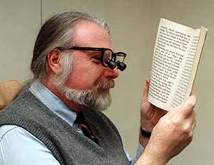


